Product
Western Blots & Immunoassays
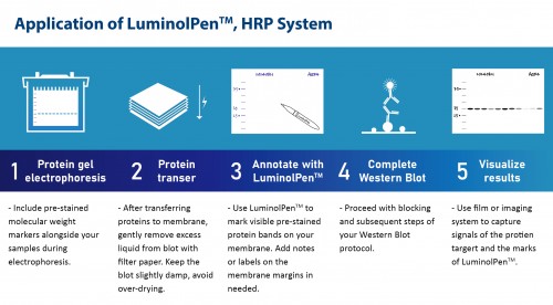
The LuminolPen™ HRP System is a chemiluminescent marker pen designed for precise data management in Western blotting. Visible blue ink reacts with ECL substrates, allowing annotations directly on PVDF or NC membranes. Use it to mark pre-stained protein positions, experiment conditions, and operation dates before blocking. Simplify your process with clear, effective documentation.
Highlights:
- Unique: the world's first luminescent color development marker pen
- Help you to evaluate the efficacy of your ECL substrate
- Data management: able to make notes and show on the image with the data result
- Compatible: applicable with most brands of ECL substrate
Order Information:
| Cat. No. | Product Name | Description |
| LH03-50 | LuminolPen™, HRP System |
About 1,000 membrane drawing |
| LH03-10 | LuminolPen™, HRP System |
About 100 membrane drawing |


Product Detail :
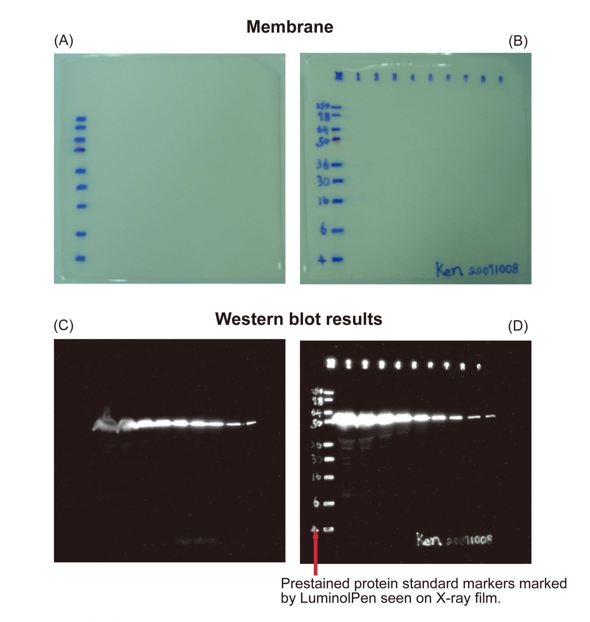
Figure 1. Labeling molecular weight and note on the membrane with LuminolPen™.
(A) and (C) are general Western blot chemiluminescence detection image; (B) and (D) are the image using LuminolPen™ to indicate the pre-stained marker.
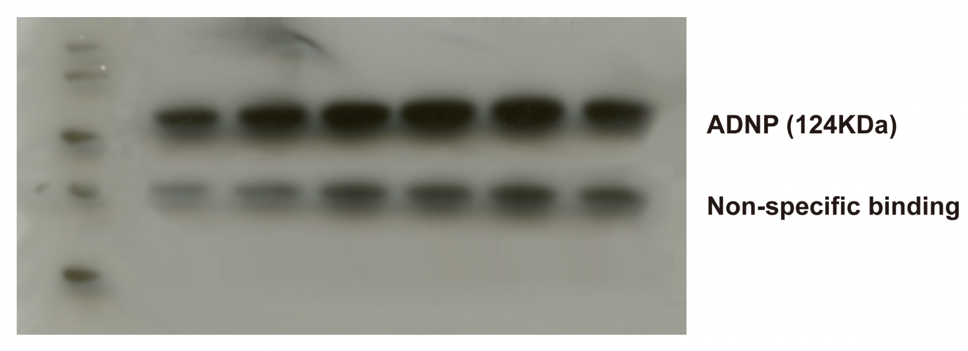
Figure 2. Distinguish target signal and non-specific binding signal with LuminolPen™
30 μg cell lysate SP-1 and detect with anti-ADNP (mouse, 1:2,000). Secondary antibody: anti-mouse IgG-HRP (1:15,000). Membrane: Hyond™ P. Detection: Hyperfilm™ ECL. Signal exposed for 30 seconds and detected by X-ray. The molecular weight indicated with a "dot" by LuminolPen™ corresponds to 72 kDa. The correct target signal (ADNP, 124 kDa) is the upper signal, not the lower one (non-specific binding).
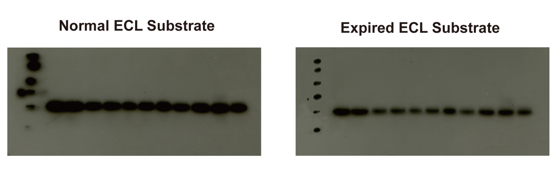
Figure 3. Evaluate ECL substrate efficacy by LuminolPen™.
30 μg cell lysate HepaG2 and detect with anti-AMP-active protein kinase (mouse, 1:1,000). Secondary antibody : anti-mouse IgG-HRP (1:10,000). Membrane: Hyond™ P. Detection: Hyperfilm™ ECL. Signal exposed for 30 seconds and detected by X-ray. The signal result present by the normal ECL substrate is stronger than using the expired ECL substrate. LuminolPen™ noted marker can be an indicator to check the result or adjust different experiments to similar experimental conditions.
Reference:
1. Tsai, Yen-Yin, et al. "Identification of the nanogold particle-induced endoplasmic reticulum stress by omic techniques and systems biology analysis." ACS nano 5.12 (2011): 9354-9369. Link
2. Chen, Han-Min, Ching-Yu Lin, and Vinchi Wang. "Amyloid P component as a plasma marker for Parkinson's disease identified by a proteomic approach." Clinical biochemistry 44.5 (2011): 377-385. Link
3. Chiu, Cheng‐Di, et al. "Investigation of the effect of hyperglycemia on intracerebral hemorrhage by proteomic approaches." Proteomics 12.1 (2012): 113-123. Link
4. Pei-Jung Katy Chung, et al. “microRNA-205 targets tight junction-related proteins during urothelial cellular differentiation.” Molecular & Cellular Proteomics (2014) 13, 9: 2321-2336 Link
5. Rahman, Md, et al. "Comprehensive Proteomic Analysis Revealed a Large Number of Newly Identified Proteins in the Small Extracellular Vesicles of Milk from Late-Stage Lactating Cows." Animals 11.9 (2021): 2506. Link
6. Rahman, Md Matiur, et al. "Identification of suitable internal control miRNAs in bovine milk small extracellular vesicles for normalization in quantitative real-time polymerase chain reaction." Membranes 13.2 (2023): 185. Link
7. Nguyen, Tan Phat, et al. "Investigation of the functional role of UNC93B1 in Nile tilapia (Oreochromis niloticus): mRNA expression, subcellular localization, and physical interaction with fish-specific TLRs." Fish & Shellfish Immunology 139 (2023): 108902. Link